Disc Herniation: What It Means and How Recovery Happens
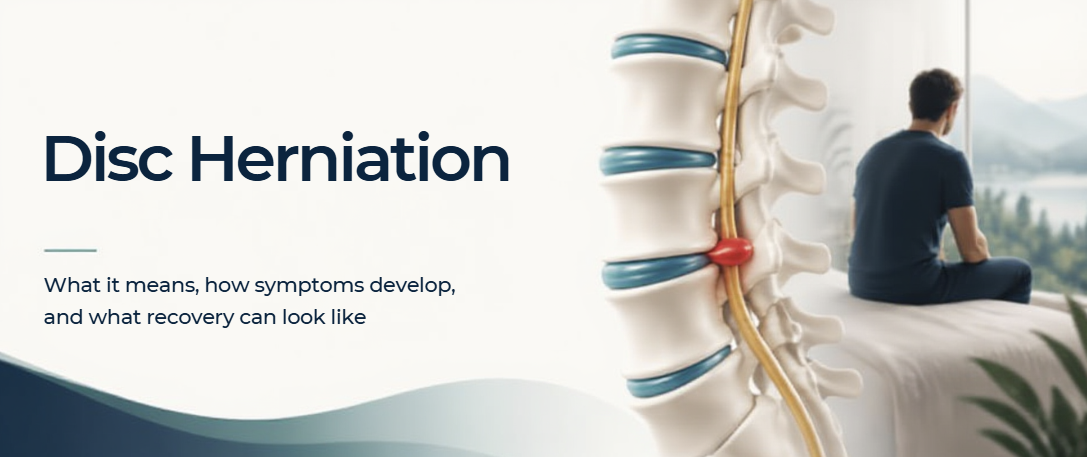
Hearing that you have a “slipped disc” can sound alarming.
It can create the impression that one of the discs in your spine has slipped out of position and needs to somehow be pushed back into place.
That is not what a disc herniation is.
A disc herniation is a change in the structure of an intervertebral disc that allows some of the inner disc material to extend beyond its usual boundary. Depending on its location and the surrounding tissues involved, it may cause back pain, irritation of a spinal nerve, pain into the leg — or sometimes no symptoms at all.
Understanding what is actually happening can make the diagnosis considerably less frightening.
What is an intervertebral disc?
Between most of the vertebrae in your spine sits an intervertebral disc.
The disc helps distribute load through the spine while allowing movement between the vertebrae.
It has two main components:
a tougher outer ring called the annulus fibrosus
a softer, gel-like central region called the nucleus pulposus
Discs are living structures that change throughout life and are influenced by factors such as ageing, genetics, loading, activity, smoking and injury.
A healthy spine is therefore not a perfectly static structure.
What is a disc herniation?
A disc herniation occurs when disc material extends beyond its normal boundary.
You may come across terms such as:
Disc bulge — the disc extends more broadly beyond its usual margin.
Disc protrusion — a more localised extension of disc material remains relatively contained.
Disc extrusion — disc material extends further through the outer layers of the disc.
Disc sequestration — a portion of the herniated material separates from the main disc.
These descriptions help clinicians and radiologists describe what the disc looks like on imaging.
But the appearance of the disc is only one part of the clinical picture.
Does a disc herniation always cause pain?
No.
Disc changes can be seen on MRI in people who have no back pain at all.
This is why an MRI should never be interpreted in isolation.
A clinician needs to ask whether the location and characteristics of the disc finding actually fit the person's symptoms and examination.
A disc herniation becomes clinically important when its location and surrounding tissue response correspond with the person's symptoms and examination findings, particularly when a nearby nerve root is irritated or compressed.
Why can a disc herniation cause leg pain?
A lumbar disc herniation can irritate or compress a nerve root travelling from the lower spine into the leg.
This can produce radicular symptoms, commonly referred to as sciatica.
Depending on which nerve is affected, symptoms may include:
pain travelling into the buttock or leg
pins and needles or tingling
numbness
altered sensation
weakness in particular muscles
changes in reflexes
The leg symptoms do not necessarily mean that something has “moved further out.”
Nerve symptoms can be influenced by both mechanical pressure and the inflammatory response around the nerve root.
This helps explain why symptoms can sometimes change considerably even though the underlying anatomy has not suddenly changed.
What about back pain?
Not every disc herniation produces sciatica.
Some people experience predominantly lower-back pain, while others have both back and leg symptoms.
Back pain is also complex. Muscles, joints, discs, connective tissues and the nervous system can all contribute to the experience of pain.
This is why finding a disc abnormality on a scan does not automatically prove that the disc is responsible for every symptom a person is experiencing.
The examination still matters.
Can a herniated disc heal?
One of the most reassuring findings from modern research is that disc herniations can change over time.
The body can gradually resorb part or, in some cases, much of the herniated material.
Research following people treated conservatively has demonstrated spontaneous resorption of lumbar disc herniations on follow-up imaging, often occurring within the first several months.
Interestingly, larger extruded and sequestrated herniations may actually have a greater likelihood of resorption than smaller contained bulges.
This can seem counterintuitive.
A dramatic-looking MRI does not automatically mean that the outlook is worse.
The body is not simply waiting for somebody to mechanically “put the disc back.” It has its own biological processes for responding to the herniated tissue.
Does the disc need to be pushed back into place?
No.
A disc herniation is not a bone that has slipped out of position.
Manual treatment does not physically push extruded disc material back inside the disc.
Conservative treatment instead aims to help manage the effects of the injury — reducing pain and irritation, maintaining or restoring movement, improving function and gradually rebuilding the person's ability to tolerate normal activity.
This distinction is important because it changes the goal from trying to “fix an MRI” to helping the person recover.
Why movement matters
When back or leg pain is severe, avoiding movement can feel like the safest option.
A short period of modifying aggravating activities may be appropriate during a painful acute episode, but recovery generally involves gradually returning to movement and activity.
Exercise-based treatment for lumbar disc herniation has been associated with improvements in pain, disability and quality of life.
The exact exercise programme will not be identical for everyone.
Depending on the person's presentation, rehabilitation may work on:
comfortable spinal and hip movement
trunk control
strength and endurance
gradual loading
movement confidence
tolerance for sitting, standing and walking
work or sporting demands
The goal is not to protect the spine from movement forever.
It is to progressively restore its ability to cope with movement and load.
Where can hands-on treatment and traction fit?
Manual therapy and traction may be used as part of conservative care in selected patients.
Research suggests that exercise, manipulation and traction can all contribute to reductions in pain and disability, although individual responses vary and treatment should be matched to the person's symptoms, neurological examination and stage of recovery.
These approaches do not physically push or pull the herniated disc back into place.
Their role is to help manage symptoms, improve movement and support rehabilitation while recovery progresses.
Where might photobiomodulation fit?
Photobiomodulation may also be used as an adjunct in patients with disc-related pain and radiculopathy.
Clinical research has reported improvements in pain, disability and movement when low-level laser therapy was added to conventional rehabilitation in patients with discogenic lumbar radiculopathy.
Its role is not to physically reverse the herniation, but to influence pain and inflammatory responses while movement and rehabilitation address function and recovery.
Do all disc herniations need surgery?
No.
In the absence of important neurological complications, conservative management is generally the first approach for lumbar disc herniation.
Many people improve substantially without surgery, and the herniated material itself may reduce in size over time.
Surgery can nevertheless be extremely valuable when it is appropriately indicated.
It may be considered when there is significant or progressive neurological loss, certain emergency neurological presentations, or severe persistent symptoms that have not responded adequately to appropriate conservative management.
The question is therefore not whether surgery is “good” or “bad.”
It is whether surgery is necessary for this particular presentation.
When does a disc herniation need urgent assessment?
Most disc herniations are not medical emergencies.
There are, however, symptoms that require urgent medical assessment.
These include:
new difficulty controlling the bladder or bowel
loss or marked alteration of sensation around the saddle or genital region
rapidly developing or progressive weakness in the leg
significant neurological deterioration
severe symptoms associated with other concerning signs such as significant trauma, fever or systemic illness
A change in bladder or bowel function together with saddle sensory changes or significant neurological symptoms can indicate cauda equina syndrome, which requires urgent medical assessment.
Progressive weakness should also not simply be treated as ordinary back pain.
What does recovery look like?
Recovery is not always a straight line.
Pain may improve before strength or nerve sensitivity has completely recovered. Some activities may become comfortable before others. Symptoms may fluctuate as activity increases.
The important question is whether the overall pattern is moving in the right direction:
Is pain becoming more manageable?
Is leg pain reducing?
Is strength stable or improving?
Can you gradually tolerate more activity?
Are you returning to normal daily, work or sporting activities?
These changes often tell us more about recovery than repeatedly focusing on the appearance of the original MRI.
The bigger picture
A disc herniation is a structural change, but it is not necessarily a permanent injury and it is not simply a disc that has slipped out of place.
Some disc herniations cause significant nerve irritation and pain. Others are found incidentally and cause no symptoms.
Many symptomatic lumbar disc herniations improve with conservative management, and research has shown that herniated disc material can naturally reduce in size over time.
Treatment therefore needs to look beyond the scan.
The location of symptoms, neurological findings, movement, strength, function and progression all help determine what the disc finding actually means for the individual person.
The goal is not simply to change what an MRI looks like. It is to restore comfortable, confident and functional movement.
References
Fardon DF, Williams AL, Dohring EJ, Murtagh FR, Rothman SLG, Sze GK. Lumbar disc nomenclature: version 2.0. Recommendations of the combined task forces of the North American Spine Society, the American Society of Spine Radiology and the American Society of Neuroradiology. The Spine Journal. 2014;14(11):2525–2545.
Brinjikji W, Luetmer PH, Comstock B, et al. Systematic literature review of imaging features of spinal degeneration in asymptomatic populations. American Journal of Neuroradiology. 2015;36(4):811–816.
Brinjikji W, Diehn FE, Jarvik JG, et al. MRI findings of disc degeneration are more prevalent in adults with low back pain than in asymptomatic controls: a systematic review and meta-analysis. American Journal of Neuroradiology. 2015;36(12):2394–2399.
Mulleman D, Mammou S, Griffoul I, Watier H, Goupille P. The role of inflammation in disk herniation-associated radiculopathy. Seminars in Arthritis and Rheumatism. 2006;35(5):267–277.
Zou T, Liu XY, Wang PC, et al. Incidence of spontaneous resorption of lumbar disc herniation: a meta-analysis. Clinical Spine Surgery. 2024;37(6):256–269.
Yaman O, Guchkha A, Vaishya S, et al. The role of conservative treatment in lumbar disc herniations: WFNS Spine Committee recommendations. World Neurosurgery: X. 2024;22:100277.
Du S, Cui Z, Peng S, et al. Clinical efficacy of exercise therapy for lumbar disc herniation: a systematic review and meta-analysis of randomized controlled trials. Frontiers in Medicine. 2025;12:1531637.
Thavarajasingam SG, et al. Exercise, manipulation and traction physiotherapy in the conservative management of lumbar disc herniation: a systematic review and meta-analysis. Brain & Spine. 2025.
Ahmed I, Gilani SA, Ahmad A, Zaidi F. Effectiveness of low-level laser therapy in patients with discogenic lumbar radiculopathy: a double-blind randomized controlled trial. Journal of Healthcare Engineering. 2022;2022:6437523.
Zileli M, et al. Role of surgery in primary lumbar disk herniation: WFNS Spine Committee recommendations. World Neurosurgery: X. 2024;22:100276.
Mustafa MA, Richardson GE, Gillespie CS, et al. Definition and surgical timing in cauda equina syndrome: an updated systematic review. PLOS ONE. 2023;18(5).
This article is intended for general educational purposes and does not replace individual medical assessment, diagnosis or treatment.